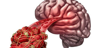
Patients suffering from stroke will now have less brain damage following the introduction of a new, less invasive, procedure that can remove a blood clot from the brain.
The Aga Khan University Hospital in Nairobi is currently the only hospital offering this procedure in the region. Strokes are a leading cause of long-lasting injury, disability, and death. The technique allows neurosurgeons to access the brain using a catheter inserted through the femoral artery in the groin to remove a clot.
This is more effective in restoring blood flow to the brain than using blood thinner medication. About 16,000 stroke cases occur each year in Kenya, of which 80 per cent are ischaemic. Most of these strokes are caused by blockage which causes lack of blood flow to parts of the brain causing damage.
When a patient has a blockage that interferes with blood flow to the brain, he is suffering from an ischaemic stroke as opposed to the hemorrhagic variety where there is bleeding into the brain.
Because blood is not flowing to the brain, cells begin to die and depending on the area affected a person may lose ability to move one side of the body, to speak, to see normally, or a number of other body functions. A person’s long term outcome depends on how much the brain is damaged and how quickly treatment begins.
For people with an ischaemic stroke, the goal of treatment is to restore blood flow to the affected area of the brain as quickly as possible, which means within six hours after the stroke begins.
The window for performing this procedure is 12 hours after which it will be of no benefit as the brain will have suffered irreversible damage. The first six hours are crucial. If the patient is rushed to our hospital immediately after the first symptoms of stroke, we are able to quickly determine the kind of stroke he is experiencing and decide if he can benefit from this procedure.
Time is of the essence because the faster the clot is removed, the sooner the patient can begin to recover normal brain functions.
The procedure involves placing a sheath in the femoral artery in the groin through which a catheter is inserted.
A special mesh is then pushed through the catheter then expanded to attach itself to the clot and the surgeon gently pulls out the clot. This takes about 30 to 45 minutes. An angiogram is carried out immediately to confirm that blood flow has been restored.
Within hours of the removal of the clot, there is improvement. A 72-year-old woman who suffered a stroke recently and was brought to the hospital was discharged after one week with medication and she is progressing well.
We were able to stop further brain damage because we managed to perform the procedure within six hours of the initiation of the stroke. Many other patients are referred to us too late to benefit from this procedure.
RESTORES FUNCTIONS
It is therefore very important to recognise stroke-related symptoms as quickly as possible to enable the surgeon time to save the patient. Common symptoms include a sudden numbness, or weakness on any limb, facial drooping, difficulty in speaking or understanding speech, sudden difficulty in walking, dizziness, loss of balance or coordination, trouble seeing with one or both eyes, severe headache and drowsiness.
This procedure, if carried out within 12 hours, successfully restores functions to the affected areas and is cost-effective, with a shorter stay in hospital.
If a patient suffers a stroke do not waste time, they require immediate care in a hospital that can coordinate emergency services, with a neurologist, intensive care services, and brain and vascular imaging with CT, or MRI scans.
Unlock a world of exclusive content today!Unlock a world of exclusive content today!